
The joints
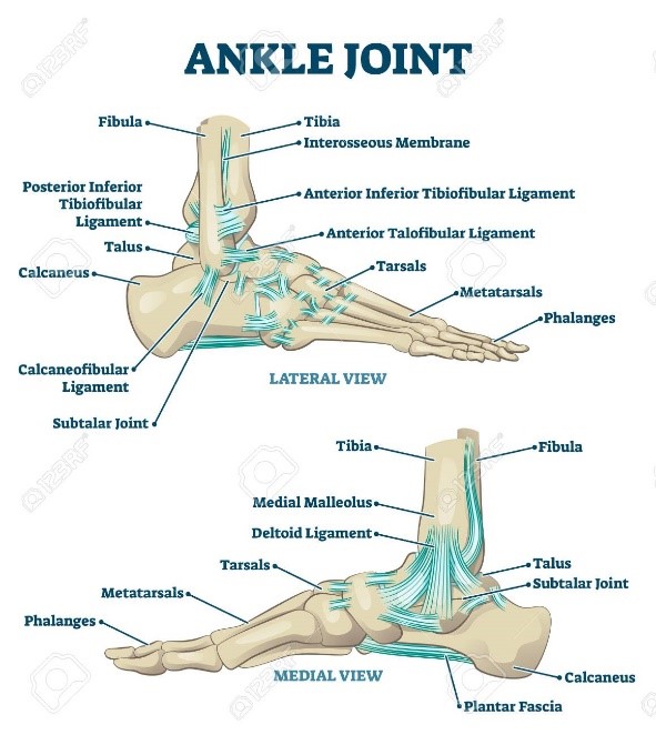
The ankle joint is a hinge joint formed by the:
- tibia
- fibula (shin bones)
- talus
As the ankle is a hinge joint, it allows an up (dorsiflexion) and down (plantarflexion) movement.
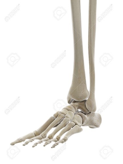
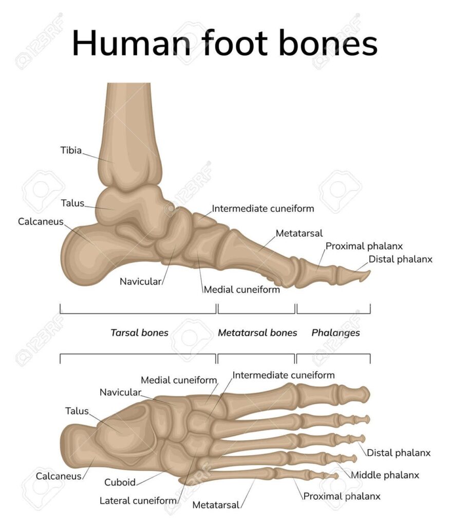
The foot is made up of 26 bones:
- 7 tarsal bones (rear foot)
- 5 metatarsals (midfoot)
- 14 phalanges (toes)
The main movement at the rear foot between the calcaneus (heel) and talus bones is in (inversion) and out (eversion).
The phalanges (toes) are hinge joints which allows for up (extension) and down (flexion) movement.
The ligaments
Ligaments are structures that connect bone to bone. They help to:
- create stability
- give the foot and ankle awareness of its position in space (proprioception)
There are many ligaments within the foot and ankle. The main ankle ligaments include:
- the lateral ligaments (on the outside of the ankle) which resist over-inversion
- the medial ligaments (on the inside of the ankle) which resist over-eversion
The muscles
The main muscles of the foot and ankle consists of 4 groups that work to move the foot:
- up (dorsiflexion)
- down (plantarflexion)
- in (inversion)
- out (eversion)
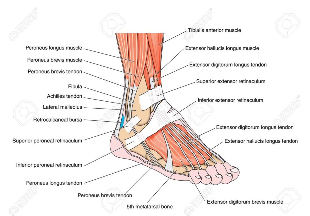
The muscles at the front of the ankle include the anterior tibial muscles. These shorten to pull the foot up and lengthen to allow to foot to point down.
The muscles at the back of the lower leg blend into the Achilles tendon. This is the largest tendon in the body and connects the calf muscles to the heel bone.
The posterior tibial muscle at the medial (inside) ankle acts to move the foot in towards the other (invert), while the peroneal muscles at the lateral (outside) ankle act to move the foot away from the other (evert).
The fascia
The fascia is a strong band of tissue on the sole of the foot also called the plantar fascia. It is thick in the middle and thinner at the sides, fanning out in a triangle shape from the heel bone (calcaneus) to the toes.
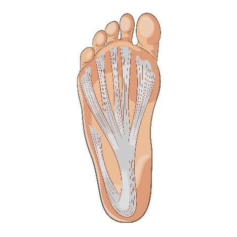
This structure is important:
- for maintaining the arch of the medial (inside) foot
- for protection of deeper structures in the foot like blood vessels and nerves
- as a shock absorber when walking